

DirectaShips in 3 business days
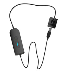
Directa TrollByte Kimera BIO Image Plate Holders · Blue · #0
SKU 107-0596MFR# 1576125233
Holder
Blue
The Directa TrollByte Kimera BIO Image Plate Yellow #0 is a miscellaneous product supplied by Directa for dental use. Catalog code 1576333053.
Directa TrollByte Kimera BIO Image Plate Holders is a sensors & imaging plate from Directa, part of the Trollbyte line.
This page carries 5 holder options: Kit, Blue, Red, Yellow, Endo. This page carries 3 plate size options: #0, #1, #2.
Find it on our Imaging & X-Ray shelf, under Sensors & Plates.
Orders placed before 3 PM ET ship the same day when the item is on our shelf (otherwise within 3 business days), and ground shipping is free over $10.
Product
- Manufacturer
- Directa
- Manufacturer #
- 1576125233
- Item code
- 107-0596
Catalog path
- Marketplace
- Clinical
- Category
- Imaging & X-Ray
- Subcategory
- Sensors & Plates
Ordering
- Status
- Ships in 3 business days
More from Sensors & Plates
Stocked in the same subcategory.


Questions
What is Trollbyte used for?
The Directa TrollByte Kimera BIO Image Plate Yellow #0 is a miscellaneous product supplied by Directa for dental use.
Which holder and plate size options are on this page?
This page carries 5 holder options: Kit, Blue, Red, Yellow, Endo. This page carries 3 plate size options: #0, #1, #2. Pick one to see its price, part number and stock.
When does it ship?
In-stock orders placed before 3 PM ET ship the same day; anything not on our shelf ships within 3 business days.Ground shipping is free over $10.
About sensors and imaging plates
This is the receptor itself, in its two forms: the solid-state sensor on a cable and the phosphor plate that is read in a scanner. Plates carry a detection layer of photostimulable crystals that absorb and temporarily store the x-ray energy until the plate is read. Plates come in the same shapes and dimensions as conventional film, though they are more rigid than film.
An intraoral digital system records the image on either a solid-state sensor or a photostimulable phosphor plate. Solid-state sensors are built around a charge-coupled device or a complementary metal-oxide semiconductor chip, often with a scintillation layer to catch the x-rays. Sensor systems are called direct: the chip sends a signal to the computer and the image appears on the monitor within seconds. Plate systems are called indirect: the exposed plate holds a latent image, is then scanned, and the scanner sends the image to the computer.
The two have different trade-offs: sensors image faster but have a relatively smaller active surface for their size, while plates are more flexible and comfortable yet more open to damage from scratches, bite marks and ambient light, and have to be scanned before the image can be read. Plates are also distinguished from sensors by a thin structure, flexibility and the absence of a cable. A report on 150 intraoral digital systems found 105 sensor-based and 45 plate-based, with 76.2 percent of the sensor systems using CMOS technology.
What to look at when choosing a sensor or plate
Size: standard receptor dimensions are 22 by 35 mm for size 0, 24 by 40 mm for size 1, 31 by 41 mm for size 2, 27 by 54 mm for size 3 and 57 by 75 mm for size 4, and size 4 exists only on plate systems. Seven plate sizes have been identified — 0, 1, 2, 3, 4, 4C and 5 — with 4C being two size 3 plates fused for occlusal imaging.
Resolution: spatial resolution is given in line pairs per millimetre, and sensor systems typically quote higher theoretical resolutions. Across the plate systems reported, pixel sizes ran from 12.5 to 64 micrometres.
Comfort and time: in a clinical comparison the total time for a full-mouth series was significantly lower with a CMOS sensor than with plates, while the patient comfort scores for the two did not differ significantly. A disadvantage named for sensors is their thickness: they are bulky and rigid, which may cause discomfort.
Life: the longevity of a plate is not yet defined, and one study states that the receptor should be replaced in cases of bending or scratches. The same study found that excessive use slightly affected the density and contrast of the radiograph, while image noise did not differ.
Sources: Sampaio-Oliveira et al. 2025, Intraoral digital radiography: a comprehensive report on the technical specifications of current and historical systems (Imaging Sci Dent) · FDA and ADA: The Selection of Patients for Dental Radiographic Examinations (revised 2012) · Ruiz et al. 2023, Could the radiographic image quality be affected by the excessive use of the photostimulable phosphor plate? (Braz Dent J) · Clinical comparison of intraoral CMOS and PSP detectors in terms of time efficiency, patient comfort, and subjective image quality, 2022 (PMC8967495) · FDA 21 CFR 872.1905, Dental x-ray film holder
